
All About Deep Brain Stimulation (DBS)

Written by: Kristi Riker OTD, OTR/L
What is DBS?
“Deep brain stimulation (DBS) has revolutionised treatment of movement disorders over recent decades, providing remarkable relief to patients with Parkinson’s disease (PD) and other profoundly debilitating disorders” (Pycroft, 2018). DBS involves surgically implanting thin electrodes into specific brain regions. The electrodes connect to a small battery-powered device (called an implantable pulse generator) placed below the skin on the upper chest. The IPG sends electrical signals that help modulate abnormal brain activity involved in movement problems. This acts like a pacemaker for brain circuits - it smooths communication between brain regions to improve movement.
How Did DBS Begin?
Back in the 1940s-1960s, surgeons actually treated tremors by intentionally lesioning (damaging) brain tissue! In Parkinson’s, some brain circuits become excessively active and begin sending abnormal rhythmic signals. This leads to movement symptoms including tremor, rigidity, slowness, and freezing. Surgeons found that damaging a very small part of those circuits can interrupt the abnormal signals and reduce symptoms like tremor or rigidity. While these surgeries sometimes helped with the movement problems, they were risky and permanent.
In the 1980s, surgeon Alim-Louis Benabid somewhat accidentally discovered that electrical stimulation during surgery could temporarily reduce symptoms. Benabid used high-frequency stimulation to test brain areas before lesioning them — and noticed the tremor stopped without needing to destroy tissue. This insight later became the basis for deep brain stimulation. DBS technologically quickly advanced in the 1990’s and gained FDA approval for tremor treatment. It has continued to progress into an increasingly sophisticated procedure and shows promise for strong future advancement.
How Does DBS Actually Work?
The exact mechanisms of DBS are not fully understood, but scientists believe it works by interfering with pathological brain rhythms. The electrical stimulation excites and/or inhibits neurons around the implanted electrodes. Low frequency stimulation appears to excite neurons while high frequency stimulation can reduce neuronal activity. This change in firing activity is what replaces the function of a lesion (tissue damage). Though unlike a lesion, DBS is not permanent and can be adjusted.
Three main areas of the brain are typically targeted in DBS: the subthalamic nucleus, the globus pallidus internus, and the ventral intermediate nucleus.
Subthalamic Nucleus (STN): this is the most common DBS target, deep within the basal ganglia system. This area of the brain helps control movement by inhibiting unwanted movement. In PD, the STN becomes overly active and fires abnormally. This causes movement pathways to become overly inhibited which leads to rigidity, tremor, slowness, and freezing. DBS electrodes placed in the STN help reduce abnormal activity and improve movement flow. This DBS target location is often used to help with tremors, rigidity, and slowness.
Globus Pallidus Internus (GPi): this area sits deeper within the basal ganglia and is one of the major output areas for motor control. With PD, output signals become inhibitory and movement initiation becomes challenging. DBS can reduce this movement inhibition and improve multiple movement problems, especially dyskinesias. This location is more commonly picked for individuals with severe involuntary movements and speech concerns.
Ventral Intermediate Nucleus: this structure is within the thalamus, near the center of the brain. Abnormal tremor circuits pass through this region and DBS placed here can greatly help with tremors, but is not as effective for rigidity and slowness.
These areas are extremely small targets (only a few millimeters) and slight changes in placement can affect outcomes. This is why surgeons use a combination of methods and sophisticated technologies to maintain delicate precision.
DBS Today:
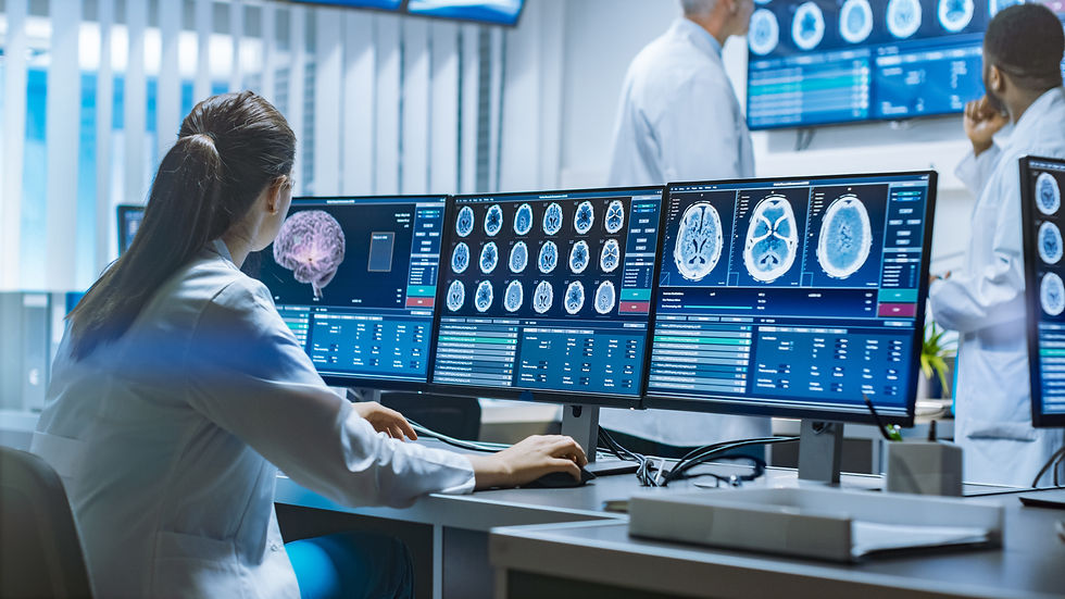
Modern technologies allow surgeons to place electrodes with millimeter precision. These methods include:
high-resolution MRI to create detailed images of the brain before surgery and locate target areas.
Stereotactic navigation is a 3D system that locates the exact spot in the brain for electrode placement (somewhat like a GPS finding the right coordinates in the brain).
Computer-guided mapping uses software to carefully guide electrode placement while avoiding blood vessels.
Microelectrode recordings to record electrical activity in the target area to ensure it is the correct location before placing the DBS electrode.
Directional leads: Older electrodes stimulated tissue in all directions. Today, electrodes have directional leads which steer electrical current more specifically to avoid affecting areas, causing side effects.
Intraoperative Testing: Sometimes patients perform movements or speak during surgery while stimulation is tested. This helps doctors check tremor reduction, speech effects, and muscle responses.
Most patients today receive:
bilateral DBS (two brain electrodes)
one implantable pulse generator (IPG) in the chest (connected to electrodes)
programmable but mostly fixed stimulation settings - doctors adjust settings during clinic visits using wireless programmers.
Surgery Risk:
Despite advanced technologies, DBS is not perfectly exact. Brain tissue can slightly shift during surgery due to tiny air or fluid changes. Researchers now think optimal DBS placement depends partly on network connectivity, not just physical anatomy. Different symptoms respond differently to slightly different regions.
The positive side - if electrodes are not placed perfectly, there are ways to compensate. Doctors can electronically “steer” stimulation after surgery through adjusting which parts of the DBS electrode send electrical signals. They can also control the strength, timing, and direction of the stimulation. This allows them to target helpful brain circuits more precisely and reduce side effects without moving the electrode.
DBS is generally considered a moderate risk brain surgery with both short-term and long-term risks
Short-term risks:
Bleeding in the brain (rare but serious, ~1–3%)
Infection of hardware (~3–5%)
Seizures (uncommon)
Confusion or temporary cognitive changes
Headache or surgical pain
Long-term risks:
Hardware infection or erosion
Lead movement (rare)
Battery replacement surgery (routine for non-rechargeable systems)
Device malfunction (uncommon)
What Does the Patient Experience?

For someone undergoing deep brain stimulation (DBS) for PD, the experience usually happens in stages over several weeks or months. The process can sound intimidating because it involves brain surgery, but many patients describe it as more structured and manageable than they expected.
Before Surgery:
DBS is usually considered when Parkinson’s symptoms such as tremor, rigidity, slowness, medication fluctuations, or dyskinesias are no longer well controlled with medication alone, but the patient still responds at least somewhat to levodopa. Before anyone is approved for DBS, they usually go through extensive testing to make sure they are a good candidate. This often includes: neurology evaluations, medication response testing, brain MRI scans, cognitive and memory testing, and mood/psychiatric screening. Doctors want to confirm that symptoms are likely to improve with DBS, cognition is stable enough, and risks are acceptable.
During surgery:
If a stereotactic frame is used, a rigid metal device is attached to the head before surgery so surgeons can navigate the brain with very high precision. When this is used, the scalp is numbed so pressure may be felt - many patients describe it as uncomfortable but tolerable. Some centers also use robotic guidance or asleep DBS techniques where imaging and computer-based navigation guide electrode placement without a rigid head frame.
Awake DBS:
Traditionally, many surgeries are done while the patient is awake but sedated. Why? Because surgeons may test stimulation in real time, monitor speech/movement, and confirm symptom improvement. Patients often report hearing conversations, feeling pressure/vibration, but not sharp pain in the brain itself (the brain has no pain receptors). Some people find awake surgery emotionally strange or tiring.
Asleep DBS
Some centers now perform DBS under general anesthesia using imaging guidance. Patients sleep through the procedure.
After surgery:
The DBS system is usually activated weeks after surgery, once swelling decreases. Initial programming appointments can be lengthy as doctors gradually adjust voltage/current, pulse width, and frequency. This phase can take weeks to months. Once settings are optimized, visits usually become much less frequent. Many patients transition to follow-up every 3–12 months depending on symptom stability, device type, and disease progression.
Long-Term Experience:
Many patients report smoother movement, reduced tremor, fewer “off” periods, and more predictable daily functioning. Over time, programming may need adjustment, medications may still be needed, and symptoms can evolve.
Most patients who benefit strongly from DBS describe the programming and recovery process as worthwhile despite the challenges.

Future DBS
Most DBS surgeries today use “open-loop” systems. This means electrical stimulation is continuous and follows programmed settings. Research is focused on use of adaptive “closed-loop” systems that can monitor abnormal brain activity and automatically adjust stimulation in real time. Researchers are developing brain-sensing implants that both stimulate and record neural activity, allowing DBS systems to respond dynamically to tremor, rigidity, or movement fluctuations. Researchers also hope future DBS devices will better treat symptoms that are currently difficult to manage, including balance problems, anxiety, sleep disturbances, and cognitive symptoms.
Future DBS research is focused on making systems more adaptive, automated, and less invasive. They are exploring noninvasive neuromodulation technologies such as focused ultrasound and other incisionless stimulation methods that may someday reduce the need for implanted hardware altogether.
Deciding on DBS
Deciding whether to undergo deep brain stimulation (DBS) for PD involves balancing potential symptom improvement against the risks and long-term commitment of brain surgery and ongoing device management.
DBS is not a cure for PD and does not stop disease progression, but it is a long-term, adjustable therapy designed to improve quality of life. Although DBS is generally considered safe in experienced centers, surgery carries moderate risks such as bleeding, infection, hardware complications, or stimulation-related side effects. Many patients who benefit from DBS report smoother movement, less tremor, and fewer “off” periods, but outcomes vary from person to person. It’s a big decision and a thoughtful team of providers is essential. Careful evaluation and realistic expectations are crucial when deciding whether DBS is the right option.
Resources:
Cavallieri F, Mulroy E, Moro E, The history of deep brain stimulation, Parkinsonism & Related Disorders, Volume 121, 2024, 105980, ISSN 1353-8020,
Eliufoo, E., Kamuyalo, C., Yusheng, T. et al. The safety profile of subthalamic nucleus and globus pallidus internus deep brain stimulation for Parkinson’s diseases: A systematic review of perioperative complications and psychological impacts. Langenbecks Arch Surg410, 131 (2025). https://doi.org/10.1007/s00423-025-03674-z
Foote A., de Waal E., Caiado F., Samman A., Ukolov A., A comprehensive review of deep brain stimulation for Parkinson's disease: The history, current state of the art and future possibilities, Medicine in Novel Technology and Devices, Volume 26, 2025, 100362, ISSN 2590-0935, https://doi.org/10.1016/j.medntd.2025.100362.
Pycroft L, Stein J, Aziz T. Deep brain stimulation: An overview of history, methods, and future developments. Brain Neurosci Adv. 2018 Dec 12;2:2398212818816017. doi: 10.1177/2398212818816017. PMID: 32166163; PMCID: PMC7058209.


Comments